Listeria L-forms: discovery of an unusual form of bacterial life
ETH Zurich researchers have discovered a new life form of Listeria monocytogenes, an opportunistic pathogen responsible for serious food poisoning. These bacteria can reproduce and proliferate as so-called L-forms. The methods to detect these bacteria should now be adapted.

For over 100 years, it was known that bacteria may lose their cell wall and can still survive. However, it was believed that this phenomenon was merely an artefact and that bacteria without cell walls do not remain viable. Recent research of a group headed by ETH Zurich Professor Martin J. Loessner, which has just been published in the high ranking journal “Molecular Microbiology” – and made it to the title page – shows that bacteria without a cell wall can be a stable form of bacterial life. Astonishingly, not only can Listeria survive without a cell wall, they are even able to reproduce and proliferate.
From cheese to the brain
Listeria (Listeria monocytogenes) are pathogens causing dangerous and often fatal cases of food-borne infections, and are frequently found in milk products such as vacherin soft cheese. The bacteria invade the human body through the epithelial cells of the intestine and spread from cell to cell., which renders them invisible to the immune system. Listeria can cross both the blood-brain barrier and the placenta barrier. Having reached the brain, they cause severe inflammation of the brain, which can be fatal. Listeria can also endanger fetuses and pregnant women.
Membrane instead of a cell wall
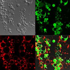
Listeria cells normally appear as small rods. If they shed their cell wall, e.g. through contact with certain antibiotics such as penicillin, they become spherical and enlarge greatly. These cell wall deficient cells are surrounded by a single membrane only. As an intermediate stage between this L-form and the rod-shaped parental cells, there is an intermediate stage from which the bacteria can rebuild their cell wall. However, once Listeria has reached the complete L-form status, there may be no way back.
The change from the normal form to the L-form is accompanied by many changes in cell metabolism and gene activity. Almost 280 of the genes of normal and L-form Listeria showed differing activity. While genes responsible for stress regulation were activated in the L-forms, genes for metabolism and energy balance were strongly repressed. The researchers interpret this as the bacterial response and active adaptation to its new lifestyle. Loessner says “L-form Listeria really have a very stressful life.”
“Culturing” the L-forms of bacteria is not easy. They need to be “bred” in a liquid medium and do not normally form colonies, so plating on a petri dish is not possible. Although L-form Listeria cells are capable of reproducing themselves, this can take time: formation of a visible colony within tubes containing a soft medium takes at least six days, compared to 16 to 20 hours for normal cells.
A new mechanism of division
The researchers were amazed by their observations on how mother cells produce L-form daughter cells. First, new vesicles form inside the large L-form cells. When these are large enough, the mother cell bursts and releases the daughter cells. At this point, these have the full genetic make-up of the mother cell, but it is still unclear how the genetic material is transferred. Interestingly, their metabolism does not start up until they have been released from the mother cell.
L-forms can grow in milk
The researchers had a reason for investigating this strange form of bacterial life: a large epidemic of Listeria with many fatalities in the US about 20 years ago. Although it is clear that this was the result of the consumption of contaminated milk, and the pathogens could be detected both on the farm from which the milk originated and in the patients who had consumed the milk, Laboratories were unable to find Listeria in the milk itself. One possible explanation is that the bacteria had been present in the milk in their reversible L-form and had thus been undetectable. Loessner says, “This is because the L-form can reproduce in milk just as well as under laboratory conditions.”
L-form Listeria can also outwit the immune system. Although macrophages, i.e. phagocytes, ingest the spherules, they seem unable to kill them in a timely fashion. While normal Listeria cells are killed after about 30 minutes, the L-forms can survive for much longer inside a macrophage. The ETH Zurich professor feels that “the immune system may have a problem if macrophages cannot recognise the L-forms as a pathogen.”
Pathologists sometimes reported small bubble-shaped
objects in brain sections from animals that had died of listeriosis, but it has
hitherto been impossible to classify these properly. Loessner hypothesizes that
these could also have involved L-form Listeria.
Reference
Dell'Era S, Buchrieser C, Couvé E, Schnell B, Briers Y, Schuppler M, Loessner MJ. Listeria monocytogenes L-forms respond to cell wall deficiency by modifying gene expression and the mode of division. Molecular Microbiology 73:306-322. Published Online: Jun 23 2009. doi:10.1111/j.1365-2958.2009.06774.x


READER COMMENTS